


All Rights for the images reserved to mugley photostream from flickr, if you keep them,please keep the credit.
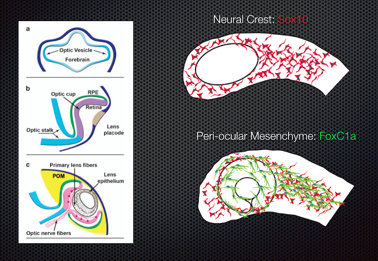
Periocular Mesenchyme
Periocular Mesenchyme (POM) are a subset of neural crest cells that ultimately generate the anterior structures of the eye, including the iris, sclera, ciliary muscles, cornea, and the trebecular mesh network.. These cells migrate along the neural crest until they encounter the retina, at which point they become strictly associated with the anterior segment region of the retina. We can visualize POM cells as they migrate to the anterior segment using a zebrafish transgenic line, FoxC1a:GFP. In contrast, we can see neural crest cell migration using the Sox10:GFP transgenic line.
While we know the ultimate purpose of the POM, we know very little of the mechanisms regulating their differentiation, migration or function. The anterior structures that arise from the POM are crucial for the function of the retina and their malfunction is associated with several ocular distrophies, including Glaucoma. However, the molecular mechanisms regulating POM differentiation and or function are largely unknown. In our lab we are therefore interested in understanding some very simple aspects of POM biology.
1) how do neural crest cells decide to become POM
2) how are POM targeted to the anterior segment
3) how do POM differentiate to form the various structures within the anterior segment
Our approach to answering these questions will involve long term time-lapse microscopy using transgenic zebrafish embryos, genomic manipulation to generate mutants and conditional knockouts for POM regulatory genes, and expression profiling of migrating POM cells.