PI: Jakub Famulski, PhD.
I was trained as a molecular cell biologist during my PhD, with a major focus on live cell imaging during cellular division. My postdoctoral training involved analysis of granule neuron progenitor differentiation and migration during cerebellar development. Currently, I am applying the zebrafish model system to study and model human ocular disease as well as analyze retinal morphogenesis. In particular I focus on colobomata, anterior segment dysgenesis and cone-rod dystrophy. In my lab we apply cutting edge confocal microscopy, transcriptomics and genome editing techniques to better understand these disorders.
Laboratory
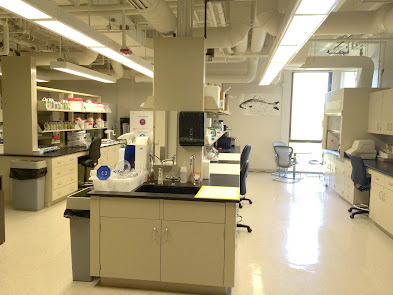
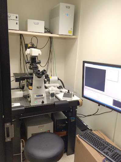
The lab is located in the TH Morgan Building in the heart of the UK campus. It is equipped with a brand new Nikon C2+ confocal system adapted specifically for long term zebrafish embryo imaging. We also have access to the Z.1 light sheet microscope for high resolution 3D live imaging. The main lab is equipped with all brand new models of molecular biology equipment including a Epoch nano spec, HRM rtPCR system, PCR machines, gel doc system and Nikon stereo microscopes. Our brand new fish facility is located across the street and houses numerous mutant, transgenic and model zebrafish lines. With plenty of space and natural light, the laboratory is inviting, efficient and professional.
Funding
2018-2024: NIH-NEI R01.
R01 EY027805-01A1
Retina Research Foundation: 2024-2025


All Rights for the images reserved to mugley photostream from flickr, if you keep them,please keep the credit.